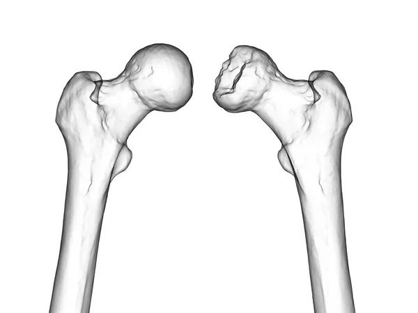
There biomedical research crosses new frontiers with the emergence of femora 3D printed, offering unprecedented innovation potential for biomechanical studies. This technique makes it possible to create realistic models at a lower cost, thus facilitating the preparation of surgical interventions and the exploration of new treatment methods. With the ability to faithfully reproduce the mechanical characteristics of the human femur, researchers plan to radically transform the way we approach orthopedic surgery and therapeutic development.
UT Southwestern researchers have developed a revolutionary 3D printing technique to create realistic human femur models. Through the use of polylactic acid, an affordable and biodegradable material, this method facilitates access to biomechanical studies while reducing costs. The tests carried out demonstrated that the 3D printed femurs display mechanical properties comparable to those of human femurs. This advance opens the way to clinical applications, such as the personalization of surgical treatments, thus making research orthopedics more accessible and effective.

Table of Contents
Toggle3D printed femurs: a revolution in biomechanics
Recently, researchers at the University of Texas at Southwestern developed an innovative technique for3D printing to create realistic human femur models. This process, which is based on the use of affordable materials, could facilitate and reduce the costs of biomechanical studies while providing reliable results. The current approach, which relies on the use of cadaver models or synthetic materials, poses many challenges such as availability and viability, which 3D printing could overcome. By integrating mechanical engineers In the process, the research opens up fascinating perspectives for the scientific community.
The impact of 3D models on biomechanics research
THE 3D printed femurs are designed from polylactic acid, a biodegradable material that reproduces the mechanical properties of human bones. By carrying out tests, the researchers were able to compare the results with the biomechanical responses of natural femurs. This discovery is significant, because it paves the way for more adapted and personalized studies and surgical practices, particularly for pathologies such asosteoporosis or traumatic fractures. The reduced costs, around $7 per femur, also represent a major advantage for researchers and medical clinics.
Towards an evolution of orthopedic surgery
With this technology, the potential applications are enormous. Doctors will be able to print models of specific pathologies to patients, thus facilitating the preparation of surgical interventions. Thanks to these realistic models, practitioners will have the opportunity to experiment with techniques before carrying out operations on patients. In this way, the future of orthopedic surgery could see extensive customizations, offering solutions tailored to each patient while preserving essential biomechanical functions.


